Encondroma
What's that?
Enchondroma (en-kon-DRO-ma) is a type of benign (noncancerous) tumor that begins in the cartilage found inside the bones. Enchondromas rarely cause pain or other symptoms, so most remain undiagnosed until x-rays are taken for an unrelated injury or condition.
In the majority of cases, enchondromas do not require treatment. In rare cases, however, multiple tumors may weaken the bone, causing it to fracture. When this occurs, surgery may be needed to remove the tumor and prevent additional fractures.
Description
Enchondromas can occur in anyone but are most common in patients between 10 and 20 years old. They are most often found in the small bones of the hand. In fact, enchondroma is the most common tumor in the hand. Enchondomas can also develop in the body's long bones, such as the femur (thighbone), tibia (shinbone), and humerus (upper arm bone).
Enchondromas are most often solitary tumors. In rare cases, however, multiple tumors can appear as part of a condition such as Ollier's disease or Maffucci's syndrome.
Single enchondromas rarely become cancerous, though the chances are a little higher in patients with Ollier's disease and Maffucci's syndrome. When enchonromas do become cancerous, they usually become a type of malignant cartilage tumor called a chondrosarcoma.
Distinguishing between a noncancerous enchondroma and the very low-grade form of a cancerous tumor can be difficult, even for orthopaedic tumor surgeons.
Cause
The exact cause of enchondromas is unknown. Some research indicates that they may result when cells turn into cartilage instead of bone.
It is not believed that the tumors are caused by radiation or chemical exposure or by any specific activity.
Symptoms
In most cases, enchondromas are not painful and do not cause any symptoms. However, if the tumors appear in the hands or feet, or if there are multiple lesions, the bone can weaken and become deformed. This can lead to pathologic bone fractures and enlargement of the affected fingers.
In patients with Ollier's disease and Maffucci's syndrome, bone deformities can be quite severe. If pain from other sources has been excluded, your doctor will carefully study the tumor to determine whether it could actually be a low-grade chondrosarcoma. Pain at night or at rest is more likely to indicate a malignant tumor. However, because pain is a common symptom of many conditions and injuries, your doctor will conduct a thorough evaluation.
Doctor Examination
Because they do not often cause symptoms, most enchondromas are found when routine x-rays are taken for another reason such as an injury or arthritis. When this occurs, your doctor will conduct an examination and order a number of tests to confirm that your tumor is actually an enchondroma—and not a more aggressive or cancerous tumor.
Physical Examination
During the exam, your doctor will take a complete medical history and ask about your symptoms. He or she will ask if your tumor is painful and when the pain occurs. There is greater concern if the pain occurs when you are at rest or at night and does not go away. Pain caused by activity is less worrisome.
In some cases, your doctor may give you an injection into the joint near the tumor. If the injection relieves your pain, it indicates that the enchondroma is not the cause.
Tests
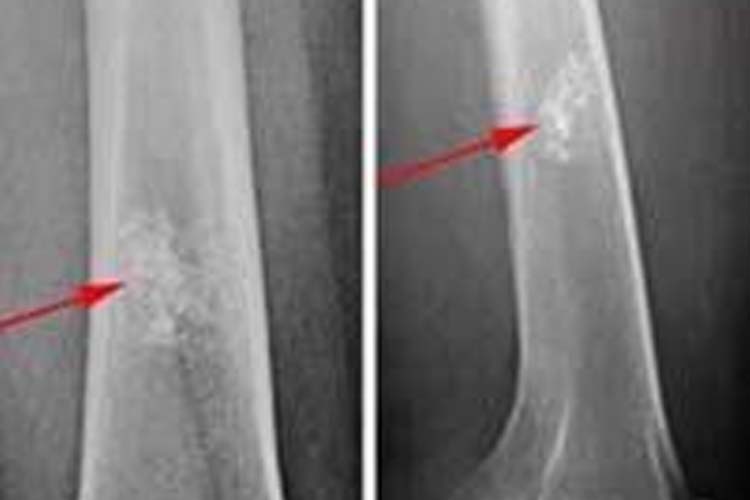
X-rays. X-rays provide images of dense structures such as bone. On x-rays, enchondromas appear as small (less than 5 cm), lobe-shaped, darkened tumors in the middle of the bone. They usually contain white spots or calcification within. The white areas of the tumor show a pattern of rings and arcs that indicates the tumor contains cartilage.
Other imaging studies. Your doctor may order a computerized tomography (CT) or magnetic resonance imaging (MRI) scan to help further evaluate your tumor. These scans give a more complete picture of the bone around the tumor. If the tumor has turned into a malignancy, the scans may show bone erosion, bone inflammation, or a mass growing outside the bone.
In some cases, your doctor may order a bone scan. During this test, a very small amount of radioactive dye is injected into the body intravenously. Both benign and malignant tumors can cause an increased uptake of the radioactive material in the bone due to bone activity. Enchondromas are typically active on bone scans.
Biopsy. A biopsy may be necessary to confirm the diagnosis of enchondroma. In a biopsy, a tissue sample of the tumor is taken and examined under a microscope.
A biopsy can be performed under local anaesthesia with a needle or as a small open operation.
Grading. The grade, or aggressiveness, of the tumor is determined by imaging studies and how the tumor looks under a microscope.
Under the microscope, enchondromas have islands of cartilage that are easy to tell apart from the normal bone that surrounds them. Usually, cartilage is not found in the center of bones. Enchondromas in the hand and foot or in patients with Ollier's disease or Maffucci's syndrome may contain more odd-looking cartilage. It may be difficult to distinguish these tumors from low-grade chondrosarcomas.
Low-grade chondrosarcomas look more cellular than enchondromas under a microscope and there is less normal bone in the tumor. Because low-grade chondrosarcomas and enchondromas look similar, experienced surgeons, radiologists, and pathologists will work together to get the best interpretation of the tumor.
Characteristics of a more aggressive tumor or a malignant chondrosarcoma include:
• Thickening of the bone's outer cortex
• Reactive bone growth on the outer surface of the bone
• Destruction of the bone by the tumor
• Soft-tissue mass
• Large amounts of bone erosion
• Bone erosion that is growing
• Erosion surrounded by reactive bone
Treatment
Nonsurgical Treatment
If your tumor does not cause symptoms, your doctor may recommend observation and monitoring to see if it grows. During this time, you may need periodic x-rays or other tests. Most doctors think that tumors without symptoms do not need to be removed.
Surgical Treatment
Curettage
Curettage is the surgical procedure most commonly used to treat enchondromas. In curettage, the tumor is scraped out of the bone. Once enchondromas are removed, most will not return. If a tumor has caused your bone to fracture, your doctor will usually allow the fracture to heal before treating the tumor. The tumor will then be curetted out to prevent another fracture.
Bone Graft
After curettage, your doctor may fill the cavity with a bone graft to stabilize the bone. A bone graft is bone taken from a donor (allograft) or from another bone in your body (autograft). In some cases, another substance may be used to fill the cavity.
Some tumors may look like simple enchondromas on x-ray—but are painful. Treatment of these lesions can be controversial. Some doctors recommend surgical curettage. Others think that the tumors are not likely to be the cause of the pain in the area—so they recommend monitoring with regular x-rays.
Unfortunately, biopsies are not often helpful in these cases. Even for specialized bone pathologists, it can be difficult to differentiate between a benign enchondroma and a low-grade chondrosarcoma. In this setting, needle biopsies are not recommended.
More aggressive tumors with bone destruction or with a mass growing outside the bone are usually chondrosarcomas. These malignant tumors need to be removed in their entirety. The specific procedure used depends upon the grade of the tumor.
Research on the Horizon
There is a great deal of ongoing research on enchondromas and chondrosarcomas. Current studies are trying to identify chemical markers that may help doctors tell the difference between benign and malignant cartilage tumors.
Other studies are looking at the different characteristics of tumors on specialized imaging studies such as MRI, CT, or PET (positron emission tomography) scans.
Mais informações em: orthoinfo.org